
Title


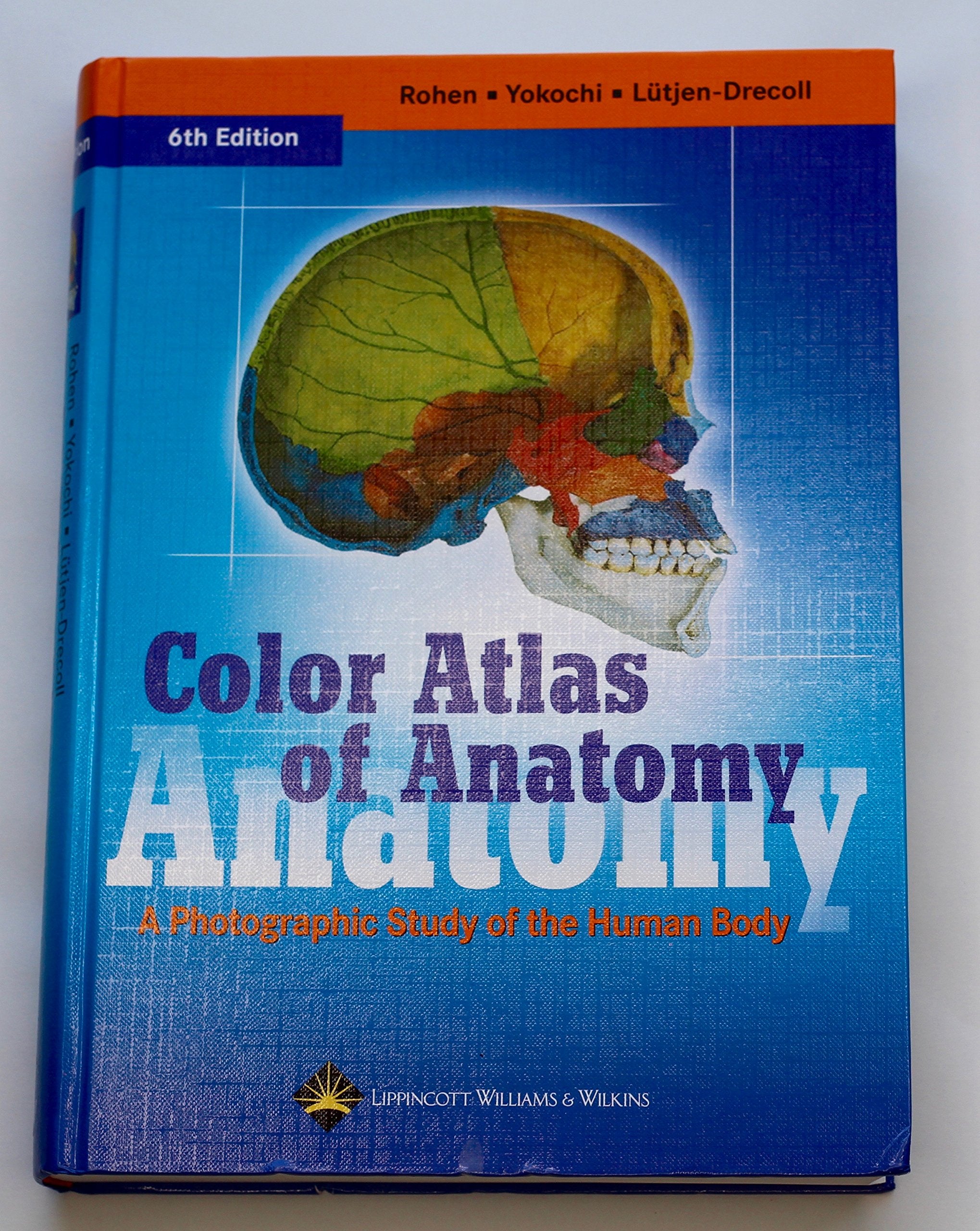
Color Atlas of Anatomy: A Photographic Study of the Human Body,Used
Sold by Ergodebooks, an authorized reseller.
Returns accepted within 30 days | support@ergodebooks.com
Shipping Information
- Free Standard Shipping — United States only
- Processing Time: 3–5 business days
- Estimated Delivery: 6–10 business days after dispatch
- Double-boxed, fully insured & discreetly packaged
- Tracking number sent via email once dispatched
Returns & Refund
Returns accepted within 30 days of delivery.
Damaged or Defective Item
Free return shipping + replacement or full refund
Wrong Item Received
Free return shipping + replacement or full refund
Change of Mind
Return shipping at customer's expense · 25% restocking fee applies
Payment Option

This atlas features outstanding fullcolor photographs of actual cadaver dissections, with accompanying schematic drawings and diagnostic images. The photographs depict anatomic structures more realistically than illustrations in traditional atlases and show students exactly what they will see in the dissection lab.Chapters are organized by region in order of a typical dissection. Each chapter presents structures both in a systemic manner from deep to surface, and in a regional manner.This edition has sixteen additional pages of clinical imagesincluding CT and MRIthat students can compare with crosssectional anatomic photographs. Many pictures have been electronically enhanced or rescanned for better contrasts.
⚠️ WARNING (California Proposition 65):
This product may contain chemicals known to the State of California to cause cancer, birth defects, or other reproductive harm.
For more information, please visit www.P65Warnings.ca.gov.
- Q: What is the main focus of the Color Atlas of Anatomy? A: The Color Atlas of Anatomy primarily focuses on providing full-color photographs of actual cadaver dissections, along with schematic drawings and diagnostic images, to help students visualize anatomical structures realistically.
- Q: How are the chapters organized in this anatomy atlas? A: Chapters in the atlas are organized by region in a typical dissection order, presenting structures both systematically from deep to surface and regionally.
- Q: What additional features does the latest edition of this atlas include? A: The latest edition includes sixteen additional pages of clinical images, including CT and MRI scans, which allow students to compare with cross-sectional anatomical photographs.
- Q: Is this anatomy atlas suitable for beginners in anatomy studies? A: Yes, the atlas is designed to assist students at various levels, including beginners, by providing clear, realistic visual representations of anatomical structures.
- Q: What type of images can I expect to find in this atlas? A: You can expect to find crisp color pictures, including drawings, photographs of cadavers, and x-ray imaging, enhancing the learning experience.
- Q: What is the binding type of the Color Atlas of Anatomy? A: The Color Atlas of Anatomy is available in hardcover binding, providing durability for frequent use.
- Q: How many pages does the Color Atlas of Anatomy contain? A: The atlas contains a total of 532 pages, offering a comprehensive resource for studying human anatomy.
- Q: Who is the author of the Color Atlas of Anatomy? A: The Color Atlas of Anatomy is authored by Johannes W. Rohen.
- Q: When was the Color Atlas of Anatomy published? A: The Color Atlas of Anatomy was published on January 1, 2006.
- Q: What condition is this book in? A: The item condition of the Color Atlas of Anatomy is rated as 'Good', indicating that it is in usable condition with some signs of wear.