
Title
Color Atlas of Histology,Used
Sold by Ergodebooks, an authorized reseller.
Returns accepted within 30 days | support@ergodebooks.com
Shipping Information
- Free Standard Shipping — United States only
- Processing Time: 3–5 business days
- Estimated Delivery: 6–10 business days after dispatch
- Double-boxed, fully insured & discreetly packaged
- Tracking number sent via email once dispatched
Returns & Refund
Returns accepted within 30 days of delivery.
Damaged or Defective Item
Free return shipping + replacement or full refund
Wrong Item Received
Free return shipping + replacement or full refund
Change of Mind
Return shipping at customer's expense · 25% restocking fee applies
Payment Option

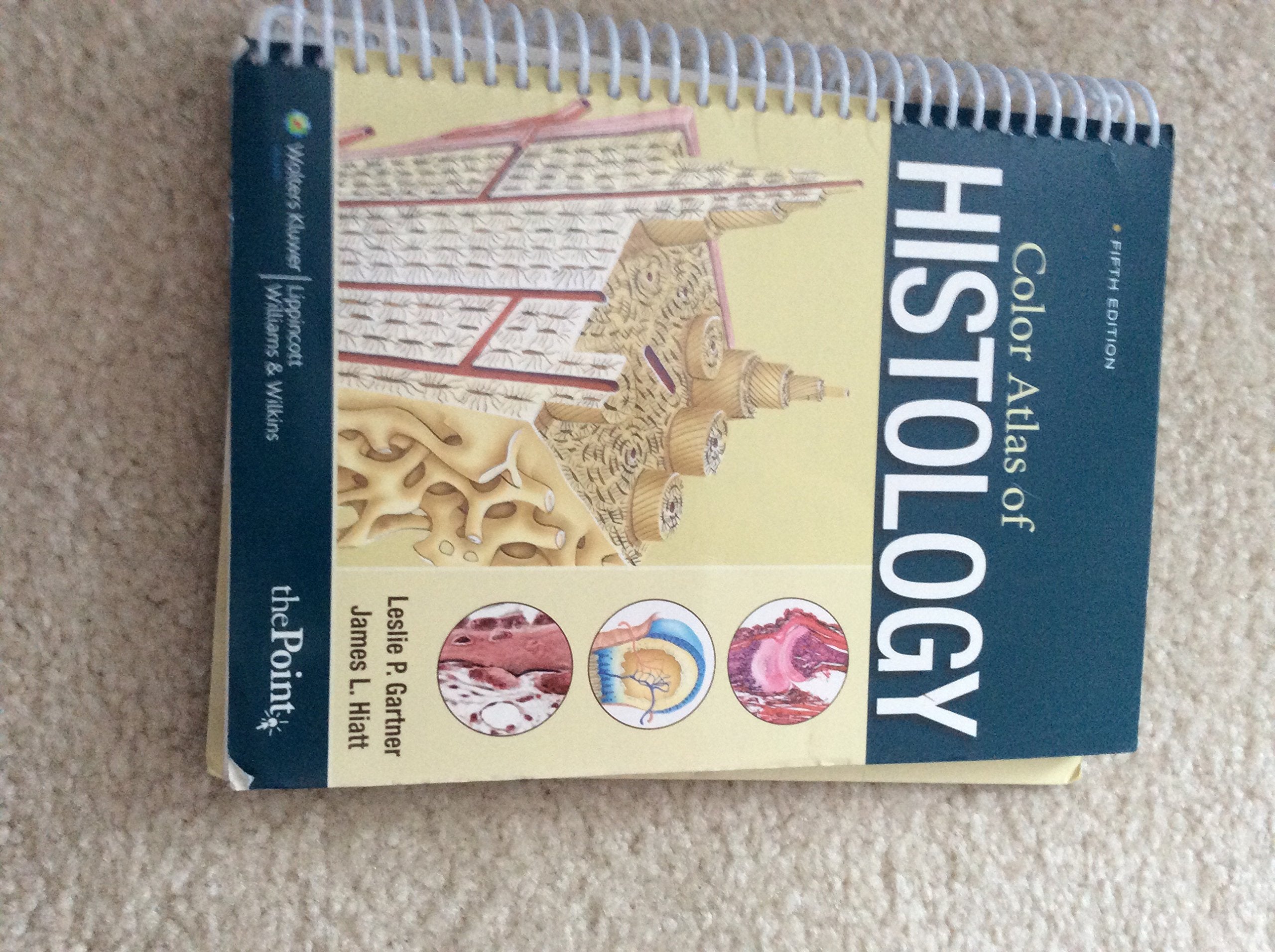
Now in its Fifth Edition, this bestselling atlas provides medical, dental, allied health, and biology students with an outstanding collection of histology images for all of the major tissue classes and body systems. This is a compact lab atlas with relevant concise text and consistent format presentation of photomicrograph plates. With a handy spiral binding that allows ease of use, it features a fullcolor art program comprising over 500 highquality photomicrographs, scanning electron micrographs, and drawings. Didactic text at the beginning of each chapter includes an Introduction, Histophysiology, Clinical Correlations, and Overview.A companion Website includes an interactive atlas and a question bank. The interactive atlas contains all the photomicrographs and electron micrographs and accompanying legends from the atlas. Images may be viewed with or without the labels and/or legends, enlarged, or compared sidebyside. A 'hotspot' feature allows students to selftest on the labeling.
⚠️ WARNING (California Proposition 65):
This product may contain chemicals known to the State of California to cause cancer, birth defects, or other reproductive harm.
For more information, please visit www.P65Warnings.ca.gov.
- Q: What is the main focus of the Color Atlas of Histology? A: The Color Atlas of Histology primarily provides medical, dental, allied health, and biology students with a comprehensive collection of histology images across major tissue classes and body systems.
- Q: How many pages does the Color Atlas of Histology have? A: The Color Atlas of Histology contains a total of 459 pages.
- Q: Is the Color Atlas of Histology suitable for beginners? A: Yes, the atlas is designed for students at various levels, including beginners, with clear didactic text and organized content to aid in understanding histology.
- Q: What type of binding does the Color Atlas of Histology have? A: The Color Atlas of Histology features a spiral binding, which allows for ease of use during lab work.
- Q: What kind of images are included in the Color Atlas of Histology? A: The atlas includes over 500 high-quality photomicrographs, scanning electron micrographs, and drawings.
- Q: Does the Color Atlas of Histology come with any online resources? A: Yes, it includes a companion website that features an interactive atlas and a question bank for additional learning support.
- Q: Who is the author of the Color Atlas of Histology? A: The Color Atlas of Histology is authored by Leslie P. Gartner.
- Q: What is the publication date of the Color Atlas of Histology? A: The Color Atlas of Histology was published on January 1, 2009.
- Q: What condition is the used book in? A: The used book is in good condition.
- Q: What is the edition of the Color Atlas of Histology? A: This is the fifth edition of the Color Atlas of Histology.