

Title
Photographic Atlas for Anatomy & Physiology, A,Used
Sold by Ergodebooks, an authorized reseller.
Returns accepted within 30 days | support@ergodebooks.com
Shipping Information
- Free Standard Shipping — United States only
- Processing Time: 3–5 business days
- Estimated Delivery: 6–10 business days after dispatch
- Double-boxed, fully insured & discreetly packaged
- Tracking number sent via email once dispatched
Returns & Refund
Returns accepted within 30 days of delivery.
Damaged or Defective Item
Free return shipping + replacement or full refund
Wrong Item Received
Free return shipping + replacement or full refund
Change of Mind
Return shipping at customer's expense · 25% restocking fee applies
Payment Option

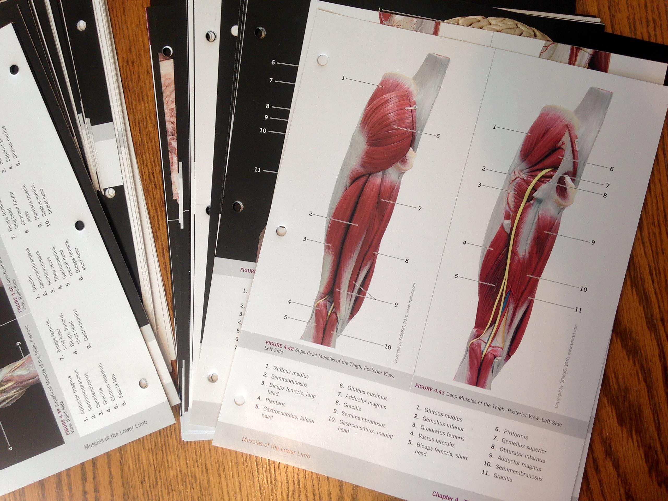
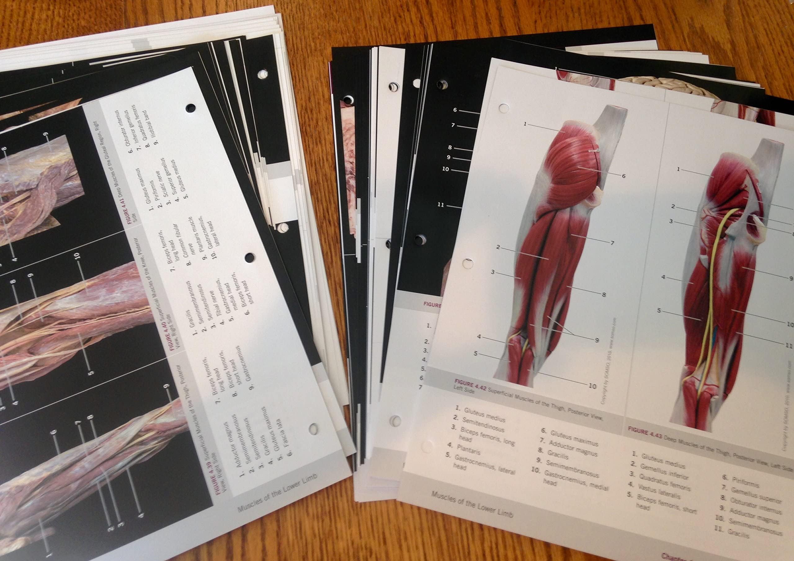
A Photographic Atlas for Anatomy & Physiology is a new visual lab study tool that helps students learn and identify key anatomical structures. Featuring photos from Practice Anatomy Lab? 3.0 and other sources, the Atlas includes over 250 cadaver dissection photos, histology photomicrographs, and cat dissection photos plus over 50 photos of anatomical models from leading manufacturers such as 3B Scientific?, SOMSO?, and DenoyerGeppert Science Company. The Atlas is composed of 13 chapters, organized by body system, and includes a final chapter with cat dissection photos. In each chapter, students will first explore gross anatomy, as seen on cadavers and anatomical models, and then conclude with relevant histological images.
⚠️ WARNING (California Proposition 65):
This product may contain chemicals known to the State of California to cause cancer, birth defects, or other reproductive harm.
For more information, please visit www.P65Warnings.ca.gov.
- Q: What is the main purpose of the Photographic Atlas for Anatomy & Physiology? A: The Photographic Atlas for Anatomy & Physiology serves as a visual lab study tool designed to help students learn and identify key anatomical structures through detailed images.
- Q: How many chapters does the atlas include? A: The atlas consists of 13 chapters organized by body system, allowing for systematic study of human anatomy.
- Q: What types of images are featured in the atlas? A: The atlas includes over 250 cadaver dissection photos, histology photomicrographs, cat dissection photos, and images of anatomical models from leading manufacturers.
- Q: Who is the author of this atlas? A: The Photographic Atlas for Anatomy & Physiology is authored by Nora Hebert.
- Q: What is the binding type of the atlas? A: The atlas is bound as a loose-leaf format, allowing for easy handling and organization of the pages.
- Q: When was the Photographic Atlas for Anatomy & Physiology published? A: The atlas was published on October 14, 2014.
- Q: What is the length of the book? A: The Photographic Atlas for Anatomy & Physiology contains a total of 240 pages.
- Q: Is this atlas suitable for beginners in anatomy? A: Yes, the atlas is designed to be a helpful resource for students at various levels, including beginners, by providing clear visual references.
- Q: Are there histological images included in the atlas? A: Yes, the atlas includes relevant histological images that complement the anatomical photographs.
- Q: Can this atlas be used for cat dissection studies? A: Yes, the atlas includes a final chapter dedicated to cat dissection photos, making it useful for veterinary anatomy studies.