
Title
Pocket Atlas of Sectional Anatomy,Used
Processing time: 1-3 days
US Orders Ships in: 3-5 days
International Orders Ships in: 8-12 days
Return Policy: 15-days return on defective items
Payment Option

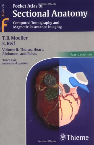
Renowned for its superb illustrations and highly practical information, the third edition of this classic reference reflects the very latest in stateoftheart imaging technology. Together with Volumes 1 and 3, this compact and portable book provides a highly specialized navigational tool for clinicians seeking to master the ability to recognize anatomical structures and accurately interpret CT and MR images. New CT and MR images of the highest quality Didactic organization using twopage units, with radiographs on one page and fullcolor illustrations on the next Concise, easytoread labeling on all figures Colorcoded, schematic diagrams that indicate the level of each section Sectional enlargements for detailed classification of the anatomical structure Comprehensive, compact, and portable, this book is ideal for use in both the classroom and clinical setting.
⚠️ WARNING (California Proposition 65):
This product may contain chemicals known to the State of California to cause cancer, birth defects, or other reproductive harm.
For more information, please visit www.P65Warnings.ca.gov.
- Q: What are the dimensions of the Pocket Atlas of Sectional Anatomy? A: The dimensions are four point four five inches by seven point five two inches by zero point five one inches. This compact size makes it portable and easy to use.
- Q: How many pages does this book contain? A: The book contains zero pages. This indicates it might be a reference or resource that is yet to be published.
- Q: What type of binding does this book have? A: The Pocket Atlas of Sectional Anatomy has a paperback binding. This design offers flexibility and ease of handling.
- Q: Who is the author of this book? A: The author is Torsten B. Moeller. He is known for his expertise in anatomical illustration and medical education.
- Q: What category does this book fall under? A: This book falls under the Medical Books category. It is especially suited for those in the medical field.
- Q: How do I use the Pocket Atlas of Sectional Anatomy effectively? A: To use this atlas effectively, read through the didactic organization that uses two-page units. Each unit has radiographs on one page and full-color illustrations on the next.
- Q: Is this book suitable for beginners in medical studies? A: Yes, this book is suitable for beginners. Its concise labeling and clear illustrations help new learners grasp anatomical concepts.
- Q: Can I use this book for clinical settings? A: Yes, this book is ideal for clinical settings. Its compact design and high-quality images make it a practical tool for clinicians.
- Q: How should I store the Pocket Atlas of Sectional Anatomy? A: Store the book in a cool, dry place away from direct sunlight. This helps maintain its quality and prevents deterioration.
- Q: What if my Pocket Atlas of Sectional Anatomy arrives damaged? A: If the book arrives damaged, you should contact the seller for a return or exchange. Most sellers have policies for handling damaged items.
- Q: How do I clean the Pocket Atlas of Sectional Anatomy? A: To clean the book, use a soft, dry cloth to wipe the cover and pages. Avoid using water or cleaners that could damage the material.
- Q: Is there a warranty for this book? A: No, typically books do not come with a warranty. However, check the seller's return policy for any guarantees.
- Q: What makes this atlas different from other anatomical books? A: This atlas features new CT and MR images of the highest quality and a unique two-page organization. This enhances learning and image interpretation.
- Q: Is this book recommended for advanced medical professionals? A: Yes, advanced medical professionals may find this book useful. Its detailed images support advanced study and interpretation of anatomy.
- Q: What are the key features of the Pocket Atlas of Sectional Anatomy? A: Key features include color-coded diagrams, detailed labeling, and sectional enlargements for better understanding of anatomical structures. These elements facilitate both learning and practical application.